 |
|
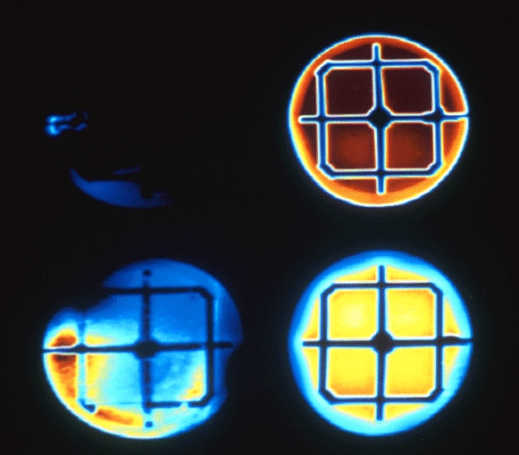
The photograph shows four individual CdS/CuInSe2 thin film solar cells(1) imaged using optical beam induced current (OBIC) with a confocal scanning beam MACROscope(2). The cells were deposited on the same substrate and connected in parallel for this expe riment. Red, yellow and blue indicates areas of high, medium and low photo-current respectively. The highest performance cell, due to its uniformity and high photo-current output, is shown on the top right corner. The other cells are progressively poore r in performance proceeding in a clockwise direction. The top left cell is almost completely dead. The size of each cell is approximately 1.5cm in diameter. Spatially resolved OBIC serves as a quality control and characterization method for solar cell.
(1) The MACROscope is a large area scanning beam imaging system that produces reflected-lignite, luminescence and OBIC images (512x5l2 pixels) with scan sizes ranging from 7x7cm down to 500x500 micro-meter (5 micro-meter resolution) in less than 5 seconds . This system was developed in the Confocal Microscopy Lab at the University of Waterloo (UW).
(2) The CdS/CuInSe2 thin film solar cells were produced by Henry Tiedje using evaporation and sputter techniques in the Thin Films Physics Lab located at UW.